- Page 1
- Page 2
- Page 3
- Page 4
- Page 5
- Page 6
- Page 7
- Page 8
- Page 9
- Page 10
- Page 11
- Page 12
- Page 13
- Page 14
- Page 15
- Page 16
- Page 17
- Page 18
- Page 19
- Page 20
- Page 21
- Page 22
- Page 23
- Page 24
- Page 25
- Page 26
- Page 27
- Page 28
- Page 29
- Page 30
- Page 31
- Page 32
- Page 33
- Page 34
- Page 35
- Page 36
- Page 37
- Page 38
- Page 39
- Page 40
- Page 41
- Page 42
- Page 43
- Page 44
- Page 45
- Page 46
- Page 47
- Page 48
- Page 49
- Page 50
- Page 51
- Page 52
- Page 53
- Page 54
- Page 55
- Page 56
- Flash version
© UniFlip.com
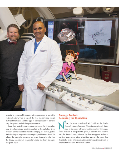
revealed a catastrophic rupture of an aneurysm in the right vertebral artery. This is one of the four major blood vessels that feed the brain, and this type of aneurysm can be particularly dangerous and challenging to control. Blood had leaked into the water system of the brain, clogging it and creating a condition called hydrocephalus. It put pressure on the brain that risked damaging the tissues, potentially leading to long-term neurological problems or death. To relieve the mounting pressure, the team inserted a tube into the brain, an external ventricular drain, to divert the cerebrospinal fluid.
Damage Control: Repairing the Dissection
ext, the team transferred Mr. Parish to the Stroke Center’s state-of-the-art Neurointerventional Suite, one of the most advanced in the country. Through a small incision in the patient’s groin, a catheter was inserted into the femoral artery. Guided by fluoroscopy—a real-time, moving image on a giant television screen—the team then threaded a series of slender catheters through the network of arteries that led into Mr. Parish’s brain.
Robert Wood Johnson I MEDICINE 7